Por favor, use este identificador para citar o enlazar este ítem:
https://repositorio.uca.edu.ar/handle/123456789/8769| Título: | IL-17 and IFN-γ expression in lymphocytes from patients with active tuberculosis correlates with the severity of the disease | Autor: | Jurado, Javier O. Pasquinelli, Virginia Álvarez, Ivana B. Peña, Delfina Rovetta, Ana I. Tateosian, Nancy L. Romeo, Horacio Musella, Rosa M. Palmero, Domingo Chuluyán, H. Eduardo García, Verónica E. |
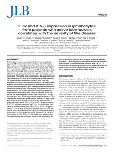
Palabras clave: | TUBERCULOSIS; ENFERMEDADES INFECCIOSAS; LINFOCITOS; CITOCINAS; CELULAS T | Fecha de publicación: | 2012 | Editorial: | Society for Leukocyte Biology | Cita: | Jurado JO, Pasquinelli V, Alvarez IB, et al. IL-17 and IFN-γ expression in lymphocytes from patients with active tuberculosis correlates with the severity of the disease. J Leukoc Biol. 2012;91(6):991–1002. doi:10.1189/jlb.1211619. Disponible en: https://repositorio.uca.edu.ar/handle/123456789/8769 | Resumen: | Abstract: Th1 lymphocytes are crucial in the immune response against Mycobacterium tuberculosis. Nevertheless, IFN-γ alone is not sufficient in the complete eradication of the bacteria, suggesting that other cytokines might be required for pathogen removal. Th17 cells have been associated with M. tuberculosis infection, but the role of IL-17-producing cells in human TB remains to be understood. Therefore, we investigated the induction and regulation of IFN-γ and IL-17 during the active disease. TB patients were classified as High and Low Responder individuals according to their T cell responses against the antigen, and cytokine expression upon M. tuberculosis stimulation was investigated in peripheral blood and pleural fluid. Afterwards, the potential correlation among the proportions of cytokine-producing cells and clinical parameters was analyzed. In TB patients, M. tuberculosis induced IFN-γ and IL-17, but in comparison with BCG-vaccinated healthy donors, IFN-γ results were reduced significantly, and IL-17 was markedly augmented. Moreover, the main source of IL-17 was represented by CD4(+)IFN-γ(+)IL-17(+) lymphocytes, a Th1/Th17 subset regulated by IFN-γ. Interestingly, the ratio of antigen-expanded CD4(+)IFN-γ(+)IL-17(+) lymphocytes, in peripheral blood and pleural fluid from TB patients, was correlated directly with clinical parameters associated with disease severity. Indeed, the highest proportion of CD4(+)IFN-γ(+)IL-17(+) cells was detected in Low Responder TB patients, individuals displaying severe pulmonary lesions, and longest length of disease evolution. Taken together, the present findings suggest that analysis of the expansion of CD4(+)IFN-γ(+)IL-17(+) T lymphocytes in peripheral blood of TB patients might be used as an indicator of the clinical outcome in active TB. | URI: | https://repositorio.uca.edu.ar/handle/123456789/8769 | ISSN: | 1938-3673 (online) | Disciplina: | MEDICINA | DOI: | 10.1189/jlb.1211619 | Derechos: | Acceso abierto | Fuente: | Journal of Leukocyte BiologyVol. 91. N° 6, 2012 |
| Aparece en las colecciones: | Artículos |
Ficheros en este ítem:
| Fichero | Descripción | Tamaño | Formato | |
|---|---|---|---|---|
| il17-ifn-expression-lymphocytes.pdf | 3,07 MB | Adobe PDF |  Visualizar/Abrir |
Visualizaciones de página(s)
329
comprobado en 09-feb-2026
Descarga(s)
368
comprobado en 09-feb-2026
Google ScholarTM
Ver en Google Scholar
Altmetric
Altmetric
Este ítem está sujeto a una Licencia Creative Commons

