
Por favor, use este identificador para citar o enlazar este ítem:
https://repositorio.uca.edu.ar/handle/123456789/8754| Título: | STED microscopy of living cells--new frontiers in membrane and neurobiology | Autor: | Eggeling, Christian Willig, Katrin I. Barrantes, Francisco José |
Palabras clave: | NEUROBIOLOGIA; MICROSCOPIA; NANOTECNOLOGIA; IMAGENES DE FLUORESCENCIA; MEMBRANA PLASMATICA | Fecha de publicación: | 2013 | Editorial: | Wiley | Cita: | Eggeling C, Willig KI, Barrantes FJ. STED microscopy of living cells – new frontiers in membrane and neurobiology. Journal of Neurochemistry. 2013;126(2):203-212. doi:10.1111/jnc.12243. Disponible en: https://repositorio.uca.edu.ar/handle/123456789/8754 | Resumen: | Abstract: Recent developments in fluorescence far-field microscopy such as STED microscopy have accomplished observation of the living cell with a spatial resolution far below the diffraction limit. Here, we briefly review the current approaches to super-resolution optical microscopy and present the implementation of STED microscopy for novel insights into live cell mechanisms, with a focus on neurobiology and plasma membrane dynamics. | URI: | https://repositorio.uca.edu.ar/handle/123456789/8754 | Disciplina: | MEDICINA | DOI: | 10.1111/jnc.12243 | Derechos: | Acceso abierto | Fuente: | Journal of Neurochemistry Vol. 126, N° 2, 2013 |
| Aparece en las colecciones: | Artículos |
Ficheros en este ítem:
| Fichero | Descripción | Tamaño | Formato | |
|---|---|---|---|---|
| sted-microscopy-living-cells.pdf | 431,68 kB | Adobe PDF | 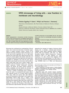 Visualizar/Abrir |
Visualizaciones de página(s)
318
comprobado en 01-jun-2026
Descarga(s)
325
comprobado en 01-jun-2026

Google ScholarTM
Ver en Google Scholar
Altmetric
Altmetric
Este ítem está sujeto a una Licencia Creative Commons
