Por favor, use este identificador para citar o enlazar este ítem:
https://repositorio.uca.edu.ar/handle/123456789/8693| Campo DC | Valor | Lengua/Idioma |
|---|---|---|
| dc.contributor.author | Ilatovskaya, Daria V. | es |
| dc.contributor.author | Blass, Gregory | es |
| dc.contributor.author | Palygin, Oleg | es |
| dc.contributor.author | Levchenko, Vladislav | es |
| dc.contributor.author | Pavlov, Tengis S. | es |
| dc.contributor.author | Grzybowski, Michael N. | es |
| dc.contributor.author | Winsor, Kristen | es |
| dc.contributor.author | Shuyskiy, Leonid S. | es |
| dc.contributor.author | Geurts, Aron M. | es |
| dc.contributor.author | Cowley, Allen W. | es |
| dc.contributor.author | Birnbaumer, Lutz | es |
| dc.contributor.author | Staruschenko, Alexander | es |
| dc.date.accessioned | 2019-09-06T17:51:22Z | - |
| dc.date.available | 2019-09-06T17:51:22Z | - |
| dc.date.issued | 2018 | - |
| dc.identifier.citation | Ilatovskaya DV, Blass G, Palygin O, et al. A NOX4/TRPC6 pathway in podocyte calcium pegulation and renal damage in diabetic kidney disease [en línea]. Journal of the American Society of Nephrology. 2018;29(7):1917-1927. doi:10.1681/ASN.2018030280 Disponible en: https://repositorio.uca.edu.ar/handle/123456789/8693 | es |
| dc.identifier.issn | 1046-6673 | - |
| dc.identifier.issn | 1533-3450 (online) | - |
| dc.identifier.uri | https://repositorio.uca.edu.ar/handle/123456789/8693 | - |
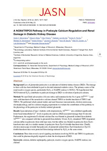
| dc.description.abstract | Abstract: Background: Loss of glomerular podocytes is an indicator of diabetic kidney disease (DKD). The damage to these cells has been attributed in part to elevated intrarenal oxidative stress. The primary source of the renal reactive oxygen species, particularly H2O2, is NADPH oxidase 4 (NOX4). We hypothesized that NOX4-derived H2O2 contributes to podocyte damage in DKD via elevation of podocyte calcium.Methods We used Dahl salt-sensitive (SS) rats with a null mutation for the Nox4 gene (SSNox4-/-) and mice with knockout of the nonselective calcium channel TRPC6 or double knockout of TRPC5 and TRPC6. We performed whole animal studies and used biosensor measurements, electron microscopy, electrophysiology, and live calcium imaging experiments to evaluate the contribution of this pathway to the physiology of the podocytes in freshly isolated glomeruli.Results Upon induction of type 1 diabetes with streptozotocin, SSNox4-/- rats exhibited significantly lower basal intracellular Ca2+ levels in podocytes and less DKD-associated damage than SS rats did. Furthermore, the angiotensin II-elicited calcium flux was blunted in glomeruli isolated from diabetic SSNox4-/- rats compared with that in glomeruli from diabetic SS rats. H2O2 stimulated TRPC-dependent calcium influx in podocytes from wild-type mice, but this influx was blunted in podocytes from Trpc6-knockout mice and, in a similar manner, in podocytes from Trpc5/6 double-knockout mice. Finally, electron microscopy revealed that podocytes of glomeruli isolated from Trpc6-knockout or Trpc5/6 double-knockout mice were protected from damage induced by H2O2 to the same extent.Conclusions These data reveal a novel signaling mechanism involving NOX4 and TRPC6 in podocytes that could be pharmacologically targeted to abate the development of DKD. | es |
| dc.format | application/pdf | - |
| dc.language.iso | eng | es |
| dc.publisher | American Society of Nephrology | es |
| dc.rights | Acceso Abierto | * |
| dc.rights.uri | http://creativecommons.org/licenses/by-nc-sa/4.0/ | * |
| dc.source | Journal of the American Society of Nephrology. 2018;29(7):1917-1927 | es |
| dc.subject | CALCIO | es |
| dc.subject | NEFROLOGIA | es |
| dc.subject | RIÑON | es |
| dc.subject | PROTEINAS | es |
| dc.subject | ENFERMEDADES RENALES | es |
| dc.subject | GENES | es |
| dc.title | A NOX4/TRPC6 pathway in podocyte calcium pegulation and renal damage in diabetic kidney disease | es |
| dc.type | Artículo | es |
| dc.identifier.doi | 10.1681/ASN.2018030280 | - |
| dc.identifier.pmid | 29793963 | - |
| uca.disciplina | MEDICINA | - |
| uca.issnrd | 1 | es |
| uca.affiliation | Fil: Ilatovskaya, Daria V. Medical College of Wisconsin. Department of Physiology; Estados Unidos | es |
| uca.affiliation | Fil: Blass, Gregory. Medical College of Wisconsin. Department of Physiology; Estados Unidos | es |
| uca.affiliation | Fil: Palygin, Oleg. Medical College of Wisconsin. Department of Physiology; Estados Unidos | es |
| uca.affiliation | Fil: Levchenko, Vladislav. Medical College of Wisconsin. Department of Physiology; Estados Unidos | es |
| uca.affiliation | Fil: Pavlov, Tengis S. Medical College of Wisconsin. Department of Physiology; Estados Unidos | es |
| uca.affiliation | Fil: Grzybowski, Michael N. Medical College of Wisconsin. Department of Physiology; Estados Unidos | es |
| uca.affiliation | Fil: Winsor, Kristen. Medical College of Wisconsin. Department of Physiology; Estados Unidos | es |
| uca.affiliation | Fil: Shuyskiy, Leonid S. Medical College of Wisconsin. Department of Physiology; Estados Unidos | es |
| uca.affiliation | Fil: Geurts, Aron M. Medical College of Wisconsin. Department of Physiology; Estados Unidos | es |
| uca.affiliation | Fil: Cowley, Allen W. Medical College of Wisconsin. Department of Physiology; Estados Unidos | es |
| uca.affiliation | Fil: Birnbaumer, Lutz. National Institute of Environmental Health Sciences. Neurobiology Laboratory; Estados Unidos | es |
| uca.affiliation | Fil: Birnbaumer, Lutz. Pontificia Universidad Católica Argentina. Facultad de Ciencias Médicas. Instituto de Investigaciones Biomédicas; Argentina | es |
| uca.affiliation | Fil: Staruschenko, Alexander. Medical College of Wisconsin. Department of Physiology; Estados Unidos | es |
| uca.version | publishedVersion | es |
| item.fulltext | With Fulltext | - |
| item.grantfulltext | open | - |
| item.languageiso639-1 | en | - |
| crisitem.author.dept | Instituto de Investigaciones Biomédicas - BIOMED | - |
| crisitem.author.dept | Laboratorio de Función y Farmacología de Canales Iónicos | - |
| crisitem.author.dept | Consejo Nacional de Investigaciones Científicas y Técnicas | - |
| crisitem.author.dept | Facultad de Ciencias Médicas | - |
| crisitem.author.orcid | 0000-0002-0775-8661 | - |
| crisitem.author.parentorg | Facultad de Ciencias Médicas | - |
| crisitem.author.parentorg | Instituto de Investigaciones Biomédicas - BIOMED | - |
| crisitem.author.parentorg | Pontificia Universidad Católica Argentina | - |
| Aparece en las colecciones: | Artículos | |
Ficheros en este ítem:
| Fichero | Descripción | Tamaño | Formato | |
|---|---|---|---|---|
| nox4-trpc6-pathway-podocyte.pdf | 1,49 MB | Adobe PDF |  Visualizar/Abrir |
Visualizaciones de página(s)
312
comprobado en 16-ene-2026
Descarga(s)
356
comprobado en 16-ene-2026
Google ScholarTM
Ver en Google Scholar
Altmetric
Altmetric
Este ítem está sujeto a una Licencia Creative Commons

