
Por favor, use este identificador para citar o enlazar este ítem:
https://repositorio.uca.edu.ar/handle/123456789/8529| Campo DC | Valor | Lengua/Idioma |
|---|---|---|
| dc.contributor.author | He, Xiju | es |
| dc.contributor.author | Li, Shoutian | es |
| dc.contributor.author | Liu, Benju | es |
| dc.contributor.author | Susperreguy, Sebastián | es |
| dc.contributor.author | Formoso, Karina | es |
| dc.contributor.author | Yao, Jinghong | es |
| dc.contributor.author | Kang, Jinsong | es |
| dc.contributor.author | Anbing, Shi | es |
| dc.contributor.author | Birnbaumer, Lutz | es |
| dc.contributor.author | Liao, Yanhong | es |
| dc.date.accessioned | 2019-08-01T01:37:29Z | - |
| dc.date.available | 2019-08-01T01:37:29Z | - |
| dc.date.issued | 2017 | - |
| dc.identifier.citation | Xiju, H., et. al. Major contribution of the 3/6/7 class of TRPC channels to myocardial ischemia/reperfusion and cellular hypoxia/reoxygenation injuries [en línea]. Proceedings of the National Academy of Sciences. 2017, 114 (23). doi:10.1073/pnas.1621384114. Disponible en: https://repositorio.uca.edu.ar/handle/123456789/8529 | es |
| dc.identifier.issn | 0027-8424 (impreso) | - |
| dc.identifier.issn | 1091-6490 (online) | - |
| dc.identifier.uri | https://repositorio.uca.edu.ar/handle/123456789/8529 | - |
| dc.identifier.uri | https://www.pnas.org/content/114/23/E4582 | - |
| dc.description.abstract | Abstract: The injury phase after myocardial infarcts occurs during reperfusion and is a consequence of calcium release from internal stores combined with calcium entry, leading to cell death by apoptopic and necrotic processes. The mechanism(s) by which calcium enters cells has(ve) not been identified. Here, we identify canonical transient receptor potential channels (TRPC) 3 and 6 as the cation channels through which most of the damaging calcium enters cells to trigger their death, and we describe mechanisms activated during the injury phase. Working in vitro with H9c2 cardiomyoblasts subjected to 9-h hypoxia followed by 6-h reoxygenation (H/R), and analyzing changes occurring in areas-at-risk (AARs) of murine hearts subjected to a 30-min ischemia followed by 24-h reperfusion (I/R) protocol, we found: (i) that blocking TRPCwith SKF96365 significantly ameliorated damage induced by H/R, including development of the mitochondrial permeability transition and proapoptotic changes in Bcl2/BAX ratios; and (ii) that AAR tissues had increased TUNEL+ cells, augmented Bcl2/BAX ratios, and increased p(S240)NFATc3, p(S473) AKT, p(S9)GSK3β, and TRPC3 and -6 proteins, consistent with activation of a positive-feedback loop in which calcium entering through TRPCs activates calcineurin-mediated NFATc3-directed transcription of TRPC genes, leading to more Ca2+ entry. All these changes were markedly reduced in mice lacking TRPC3, -6, and -7. The changes caused by I/R in AAR tissues were matched by those seen after H/R in cardiomyoblasts in all aspects except for p-AKT and p-GSK3β, which were decreased after H/R in cardiomyoblasts instead of increased. TRPC should be promising targets for pharmacologic intervention after cardiac infarcts. | es |
| dc.format | application/pdf | es |
| dc.language.iso | eng | es |
| dc.publisher | National Academy of Sciences | es |
| dc.rights | Acceso Abierto | es |
| dc.rights.uri | https://creativecommons.org/licenses/by-nc-sa/4.0/ | es |
| dc.source | Proceedings of the National Academy of Sciences 2017, 114 (23) | es |
| dc.source | ISSN 0027-8424 (impreso) | es |
| dc.source | ISSN 1091-6490 (online) | es |
| dc.subject | INFARTO DEL MIOCARDIO | es |
| dc.subject | CALCIO | es |
| dc.subject | APOPTOSIS | es |
| dc.subject | MUERTE CELULAR | es |
| dc.title | Major contribution of the 3/6/7 class of TRPC channels to myocardial ischemia/reperfusion and cellular hypoxia/reoxygenation injuries | es |
| dc.type | Artículo | es |
| uca.path | Facultad de Ciencias Médicas|Instituto de Investigaciones Biomédicas (BIOMED UCA-CONICET)|Artículos | es |
| uca.disciplina | MEDICINA | es |
| uca.filename | /home/data-uca-generic/folder_facultad/major-contribution-367-class/metadata.xml | es |
| uca.issnrd | 1 | es |
| uca.affiliation | Fil: He, Xiju. Huazhong University of Science and Technology. Tongji Medical College. Department of Anatomy; China | es |
| uca.affiliation | Fil: He, Xiju. Huazhong University of Science and Technology. Tongji Medical College. Institute of Brain Research; China | es |
| uca.affiliation | Fil: He, Xiju. Hubei University of Medicine. Department of Anatomy; China | es |
| uca.affiliation | Fil: Li, Shoutian. Huazhong University of Science and Technology. Tongji Medical College. Department of Anatomy; China | es |
| uca.affiliation | Fil: Li, Shoutian. Huazhong University of Science and Technology. Tongji Medical College. Institute of Brain Research; China | es |
| uca.affiliation | Fil: Liu, Benju. Huazhong University of Science and Technology. Tongji Medical College. Department of Anatomy; China | es |
| uca.affiliation | Fil: Liu, Benju. Huazhong University of Science and Technology. Tongji Medical College. Institute of Brain Research; China | es |
| uca.affiliation | Fil: Susperreguy, Sebastian. Pontificia Universidad Católica Argentina. Facultad de Ciencias Médicas. Instituto de Investigaciones Biomédicas; Argentina | es |
| uca.affiliation | Fil: Formoso, Karina. Pontificia Universidad Católica Argentina. Facultad de Ciencias Médicas. Instituto de Investigaciones Biomédicas; Argentina | es |
| uca.affiliation | Fil: Yao, Jinghong. Huazhong University of Science and Technology. Tongji Medical College. Union Hospital. Department of Infectious Disease; China | es |
| uca.affiliation | Fil: Kang, Jinsong. Huazhong University of Science and Technology. Tongji Medical College. Tongji Hospital. Department of Surgery; China | es |
| uca.affiliation | Fil: Anbing, Shi. Huazhong University of Science and Technology. Tongji Medical College. Department of Biochemistry and Molecular Biology; China | es |
| uca.affiliation | Fil: Birnbaumer, Lutz. Pontificia Universidad Católica Argentina. Facultad de Ciencias Médicas. Instituto de Investigaciones Biomédicas; Argentina | es |
| uca.affiliation | Fil: Birnbaumer, Lutz. Research Triangle Park. National Institute of Environmental Health Sciences. Neurobiology Laboratory; Estados Unidos | es |
| uca.affiliation | Fil: Liao, Yanhong. Huazhong University of Science and Technology. Tongji Medical College. Department of Anatomy; China | es |
| uca.affiliation | Fil: Liao, Yanhong. Huazhong University of Science and Technology. Tongji Medical College. Institute of Brain Research; China | es |
| uca.version | publishedVersion | es |
| item.fulltext | With Fulltext | - |
| item.languageiso639-1 | en | - |
| item.grantfulltext | open | - |
| crisitem.author.dept | Instituto de Investigaciones Biomédicas - BIOMED | - |
| crisitem.author.dept | Laboratorio de Función y Farmacología de Canales Iónicos | - |
| crisitem.author.dept | Consejo Nacional de Investigaciones Científicas y Técnicas | - |
| crisitem.author.dept | Facultad de Ciencias Médicas | - |
| crisitem.author.orcid | 0000-0002-0775-8661 | - |
| crisitem.author.parentorg | Facultad de Ciencias Médicas | - |
| crisitem.author.parentorg | Instituto de Investigaciones Biomédicas - BIOMED | - |
| crisitem.author.parentorg | Pontificia Universidad Católica Argentina | - |
| crisitem.author.parentorg | Facultad de Ciencias Médicas | - |
| crisitem.author.parentorg | Instituto de Investigaciones Biomédicas - BIOMED | - |
| crisitem.author.parentorg | Pontificia Universidad Católica Argentina | - |
| crisitem.author.parentorg | Facultad de Ciencias Médicas | - |
| crisitem.author.parentorg | Instituto de Investigaciones Biomédicas - BIOMED | - |
| crisitem.author.parentorg | Pontificia Universidad Católica Argentina | - |
| Aparece en las colecciones: | Artículos | |
Ficheros en este ítem:
| Fichero | Descripción | Tamaño | Formato | |
|---|---|---|---|---|
| major-contribution-367-class.pdf | 2,13 MB | Adobe PDF | 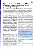 Visualizar/Abrir |
Visualizaciones de página(s)
312
comprobado en 04-jul-2026
Descarga(s)
274
comprobado en 04-jul-2026

Google ScholarTM
Ver en Google Scholar
Este ítem está sujeto a una Licencia Creative Commons
