Por favor, use este identificador para citar o enlazar este ítem:
https://repositorio.uca.edu.ar/handle/123456789/17413| Campo DC | Valor | Lengua/Idioma |
|---|---|---|
| dc.contributor.author | Caval Holme, Franklin S. | es |
| dc.contributor.author | Aranda, Marcos L. | es |
| dc.contributor.author | Chen, Andy Q. | es |
| dc.contributor.author | Tiriac, Alexandre | es |
| dc.contributor.author | Zhang, Yizhen | es |
| dc.contributor.author | Smith, Benjamin | es |
| dc.contributor.author | Birnbaumer, Lutz | es |
| dc.contributor.author | Schmidt, Tiffany M. | es |
| dc.contributor.author | Feller, Marla B. | es |
| dc.date.accessioned | 2023-11-06T23:14:45Z | - |
| dc.date.available | 2023-11-06T23:14:45Z | - |
| dc.date.issued | 2022 | - |
| dc.identifier.citation | Caval Holme, F. S. The retinal basis of light aversion in neonatal mice [en línea]. The Journal of Neuroscience. 2022, 42(20) 4101-4115. doi: 10.1523/JNEUROSCI.0151-22.2022. Disponible en: https://repositorio.uca.edu.ar/handle/123456789/17413 | es |
| dc.identifier.issn | 0270-6474 (impreso) | - |
| dc.identifier.issn | 1529-2401 (online) | - |
| dc.identifier.uri | https://repositorio.uca.edu.ar/handle/123456789/17413 | - |
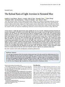
| dc.description.abstract | Aversive responses to bright light (photoaversion) require signaling from the eye to the brain. Melanopsin-expressing intrinsically photosensitive retinal ganglion cells (ipRGCs) encode absolute light intensity and are thought to provide the light signals for photoaversion. Consistent with this, neonatal mice exhibit photoaversion before the developmental onset of image vision, and melanopsin deletion abolishes photoaversion in neonates. It is not well understood how the population of ipRGCs, which constitutes multiple physiologically distinct types (denoted M1-M6 in mouse), encodes light stimuli to produce an aversive response. Here, we provide several lines of evidence that M1 ipRGCs that lack the Brn3b transcription factor drive photoaversion in neonatal mice. First, neonatal mice lacking TRPC6 and TRPC7 ion channels failed to turn away from bright light, while two photon Ca21 imaging of their acutely isolated retinas revealed reduced photosensitivity in M1 ipRGCs, but not other ipRGC types. Second, mice in which all ipRGC types except for Brn3bnegative M1 ipRGCs are ablated exhibited normal photoaversion. Third, pharmacological blockade or genetic knockout of gap junction channels expressed by ipRGCs, which reduces the light sensitivity of M2-M6 ipRGCs in the neonatal retina, had small effects on photoaversion only at the brightest light intensities. Finally, M1s were not strongly depolarized by spontaneous retinal waves, a robust source of activity in the developing retina that depolarizes all other ipRGC types. M1s therefore constitute a separate information channel between the neonatal retina and brain that could ensure behavioral responses to light but not spontaneous retinal waves... | es |
| dc.format | application/pdf | es |
| dc.language.iso | eng | es |
| dc.publisher | Society for Neuroscience | es |
| dc.rights | Acceso abierto | * |
| dc.rights.uri | http://creativecommons.org/licenses/by-nc-sa/4.0/ | * |
| dc.source | The Journal of Neuroscience. Vol.42, No.20, 4101-4115, 2022 | es |
| dc.subject | RETINA | es |
| dc.subject | DESARROLLO | es |
| dc.subject | ENUCLEACION | es |
| dc.subject | CORRIENTE FOTOELECTRICA | es |
| dc.subject | FOTOFOBIA | es |
| dc.title | The retinal basis of light aversion in neonatal mice | es |
| dc.type | Artículo | es |
| dc.identifier.doi | 10.1523/JNEUROSCI.0151-22.2022 | - |
| dc.identifier.pmid | 35396331 | - |
| uca.disciplina | MEDICINA | es |
| uca.issnrd | 1 | es |
| uca.affiliation | Fil: Caval Holme, Franklin S. University of California Berkeley. Helen Wills Neuroscience Institute; Estados Unidos | es |
| uca.affiliation | Fil: Aranda, Marcos L. Northwestern University. Department of Neurobiology; Estados Unidos | es |
| uca.affiliation | Fil: Chen, Andy Q. University of California Berkeley. Department of Molecular and Cell Biology; Estados Unidos | es |
| uca.affiliation | Fil: Tiriac, Alexandre. University of California Berkeley. Department of Molecular and Cell Biology; Estados Unidos | es |
| uca.affiliation | Fil: Zhang, Yizhen. University of California Berkeley. Department of Molecular and Cell Biology; Estados Unidos | es |
| uca.affiliation | Fil: Smith, Benjamin. University of California Berkeley. School of Optometry; Estados Unidos | es |
| uca.affiliation | Fil: Birnbaumer, Lutz. Pontificia Universidad Católica Argentina; Argentina | es |
| uca.affiliation | Fil: Birnbaumer, Lutz. National Institute of Environmental Health Sciences. National Institutes of Health. Signal Transduction Laboratory; Estados Unidos | es |
| uca.affiliation | Fil: Schmidt, Tiffany M. Northwestern University. Department of Neurobiology; Estados Unidos | es |
| uca.affiliation | Fil: Schmidt, Tiffany M. Northwestern University Feinberg School of Medicine. Department of Ophthalmology; Estados Unidos | es |
| uca.affiliation | Fil: Feller, Marla B. University of California Berkeley. Helen Wills Neuroscience Institute; Estados Unidos | es |
| uca.affiliation | Fil: Feller, Marla B. University of California Berkeley. Department of Molecular and Cell Biology; Estados Unidos | es |
| uca.version | publishedVersion | es |
| item.grantfulltext | open | - |
| item.languageiso639-1 | en | - |
| item.fulltext | With Fulltext | - |
| crisitem.author.dept | Instituto de Investigaciones Biomédicas - BIOMED | - |
| crisitem.author.dept | Laboratorio de Función y Farmacología de Canales Iónicos | - |
| crisitem.author.dept | Consejo Nacional de Investigaciones Científicas y Técnicas | - |
| crisitem.author.dept | Facultad de Ciencias Médicas | - |
| crisitem.author.orcid | 0000-0002-0775-8661 | - |
| crisitem.author.parentorg | Facultad de Ciencias Médicas | - |
| crisitem.author.parentorg | Instituto de Investigaciones Biomédicas - BIOMED | - |
| crisitem.author.parentorg | Pontificia Universidad Católica Argentina | - |
| Aparece en las colecciones: | Artículos | |
Ficheros en este ítem:
| Fichero | Descripción | Tamaño | Formato | |
|---|---|---|---|---|
| retinal-basis-light.pdf | 4,11 MB | Adobe PDF |  Visualizar/Abrir |
Visualizaciones de página(s)
51
comprobado en 27-abr-2024
Descarga(s)
13
comprobado en 27-abr-2024
Google ScholarTM
Ver en Google Scholar
Altmetric
Altmetric
Este ítem está sujeto a una Licencia Creative Commons

